


Brachytherapy
The treatment is divided into the following steps, exemplified by the liver and the lung:
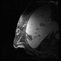
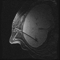
Under local anesthesia and administration of pain medication, the tumor is punctured under computer tomographic (CT), magnetic resonance imaging view with subsequent insertion of the catheters into the target volume.
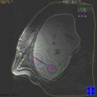
This is followed by the computer-assisted calculation of a radiation plan and computer-controlled insertion of the radiation source into the tumor via the catheters.
After completion of the irradiation, which usually lasts only a few minutes to 1 hour, the catheters are removed and hemostatic sponges are inserted into the puncture channel.
The patient is then monitored for a few more hours and transferred back to the ward. If there are no complications, the patient can be discharged on the 2nd day after the procedure.
The success of the therapy is monitored by CT and/or MRI and blood tests at 3-month intervals. For this purpose, the patient (if desired) automatically receives an appointment through our outpatient clinic.
 1
(Bild 1 von 3)
Vorwärts »
« Zurück
2
(Bild 2 von 3)
Vorwärts »
« Zurück
3
(Bild 3 von 3)
1
(Bild 1 von 3)
Vorwärts »
« Zurück
2
(Bild 2 von 3)
Vorwärts »
« Zurück
3
(Bild 3 von 3)
Images: Brachytherapy for liver metastases: first MRI-guided placement of one catheter per tumor followed by irradiation through the catheters using Iridium 192 radiation source.
Contact
If you have any further questions about the therapy, checking the indication or making an appointment, our outpatient clinic for microtherapy in the Department of Radiology and Nuclear Medicine can be contacted by phone at +49 (391) 67-13199) or by e-mail at