UNIVERSITY CLINIC FOR RADIOLOGY AND NUCLEAR MEDICINE

Selective Internal Radio Therapy (SIRT)

SIRT stands for "Selective Internal Radiotherapy" (synonym: radioembolization of the liver) and is a newer, minimally invasive form of treatment for malignant liver tumors for patients for whom surgery is not an option or/and chemotherapy has proven ineffective. SIRT is performed jointly by nuclear medicine physicians and radiologists under angiographic guidance.

How does SIRT work?

SIRT3

SIRT is based on the introduction of small radioactive beads ("microspheres", product name: SIR-Spheres® with a diameter of c. 35 µm) into the tumour tissue of the liver. After local anaesthesia, the artery in the groin is punctured and a flexible rubber valve - a so-called "catheter sheath" - is inserted into the vessel. A catheter is inserted into the hepatic artery and from there the microspheres are applied. The radioactive element yttrium-90 contained in the spheres radiates for several days (physical half-life approx. 64 hours) over a short distance of a maximum of 11 millimetres with a high local effect ("beta radiation"). This can reduce the risk of damage to surrounding, healthy organ structures.

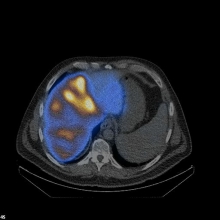
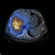
Image: Spect/low-dose CT showing tumor-oriented deposition of the 90Yttrium-labelled SIR-Spheres® in the right liver lobe in a patient with liver metastases from colorectal cancer.

When can SIRT be performed?

Indications for SIRT are primary (e.g. hepatocellular carcinoma (HCC), cholangiocellular carcinoma (CCC)) and secondary (e.g. metastases of colorectal carcinoma, breast carcinoma, melanoma, carcinoids) tumours of the liver. In advance, it must be ensured that other local and systemic therapy methods are not (or no longer) possible or do not offer sufficient treatment success. These include, for example, surgery, thermal ablation, brachytherapy or local or systemic chemotherapy.

In addition, the following criteria, among others, must be met:

  • Other therapeutic options largely exhausted
  • No evidence of reflux of accumulation into other organ areas in preparatory angiography
  • Shunt volume between liver and lung < 20%.
  • Preserved liver function
  • Leading tumor burden in the liver
What other preliminary examinations are necessary?

The intervention itself is first preceded by an examination of the indication on the basis of all available images and therapy histories. This is done after interdisciplinary consultation with the treating oncologists, surgeons, and radiotherapists, usually within the framework of an interdisciplinary tumor board.

If it turns out that SIRT treatment is indeed a promising option, further examinations are necessary for which the patients are admitted to our DR7 ward as inpatients.

During this stay, which usually lasts two to three days, an MRI of the liver and a CT of the chest and abdomen are carried out in addition to a detailed determination of laboratory values. The aim of the latter two examinations is to determine the current extent and severity of the liver disease once again at the current time.

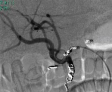
This is followed by an evaluation angiography, in which the vascular supply to the liver is made visible by means of a vascular image (angiography) under fluoroscopy. This is done - as later in the therapy - by inserting a small catheter into the groin, which is advanced to the liver region.

Vessels of the liver that show an atypical course and lead to other organs and thus represent a problem for SIRT can be detected early and closed if necessary. In addition, the actual therapy is simulated by injecting a radioactive substance with similar properties to the therapeutic microspheres. However, this substance does not contain any damaging radiation. Thus, the distribution of the actual therapeutic substance can be estimated in advance and a flow (shunt) into the lung can be excluded.

How does the actual therapy take place?

The results are then evaluated in an interdisciplinary, radiological-nuclear medicine discussion and assessed with regard to the technical feasibility of 90Yttrium radioembolisation. If there are no contraindications for carrying out the therapy, the actual treatment takes place during a renewed inpatient stay on our ward approx. 1-2 weeks after the preliminary examination. At this time, a new vascular imaging of the liver is carried out in preparation for the treatment. If necessary, small vascular branches are closed in order to exclude with a high degree of certainty any outflow of the therapy substance into other organs. Then the therapy substance is injected slowly and in portions (approx. over 30 minutes) via the catheter into the hepatic artery and under examination of the respective flow conditions.Depending on the tumour involvement of the liver, the therapy of both liver lobes is divided into 2 sessions with an interval of approx. 4 - 6 weeks in order to ensure the greatest possible protection of the healthy liver tissue. After the administration of SIR-Spheres®, the catheter is removed and, as in the pre-diagnosis, the patient should then stay in bed for approx. 24 hours to prevent possible bleeding from the groin. After the therapy, the distribution of the SIR-Spheres® is documented in a whole and partial body scintigraphy. Due to the radioactive treatment, an inpatient stay of at least 2 days is prescribed by law after the therapy, so that patients can usually be discharged after 2-3 days if the procedure is free of complications.

What are the possible side effects?

The treatment is usually well tolerated. However, some patients experience upper abdominal pain, nausea, and fever for a short time (possibly already during the treatment but also in the first 2 weeks after the therapy), but these can usually be treated very well by administering medication and usually subside quickly. Often, fatigue and reduced appetite persist for several days or weeks after treatment. Very rarely, serious side effects can occur, e.g. if, despite all precautions, microspheres are drained into other organs (e.g. the stomach, pancreas, lungs). Also, in rare cases, radiation exposure to normal liver tissue can lead to a temporary or permanent deterioration of liver function (radiation hepatitis). This effect is usually only observed about 2 months after therapy. In order to prevent such a postradiogenic liver function disorder, the patients receive liver and stomach protective medication until the second SIRT session and beyond for the next 8 weeks. This is explained to the patients in detail during their stay in the hospital. 

Is the treatment painful?

Each patient is given peripheral venous access so that infusions or medication can be administered during the procedure. In the area of the access route in the groin, the puncture site is locally anaesthetised. Under this medication, the procedure is usually painless. During the treatment, the patient's vital parameters (pulse, blood pressure) are checked at regular intervals and also documented. Occasionally, a post embolisation syndrome occurs after the procedure. Minutes to hours after the procedure, the patient may experience more or less severe upper abdominal pain. Nausea, vomiting, and fever are also observed. However, these symptoms can be controlled very well with medication. In rare cases, these symptoms can last up to a week after the therapy.

How high is the radiation exposure?

The radioactive element yttrium-90 contained in the beads radiates over several days (physical half-life approx. 64 hours) over a short distance of a maximum of 11 Millimeters range with a high local effect ("beta radiation"). This results in a very high radiation dose in the liver or in the tumor tissue, while only a very low radiation exposure occurs in the healthy liver tissue or in the surrounding organ structures.

Within the liver, interaction with the tissue produces X-ray bremsstrahlung, which has a greater range than beta radiation. Therefore, in Germany, a 48-hour stay in an appropriately equipped ward is required.

After approx. 11 days, no radiation emission from the patient is detectable. The radiation emitted by the patient is so low that after the first 48 hours no further protective measures against other persons are necessary.

Only pregnant women should be kept away from SIRT in the first few days.

What kind of results can be expected?

Treatment with SIRT cannot cure the tumor disease, even if the treatment is able to reduce the tumor in individual patients to such an extent that it is no longer detectable with the usual examination methods. The primary goal of treatment is to prolong life and improve quality of life. In individual cases, however, SIRT can achieve such a strong reduction in tumor volume that, in the course of treatment, the remaining parts of the tumor can be surgically removed or treated by microwave ablation/brachytherapy. In most cases, a decrease in tumor markers is detectable as a sign of the therapy's success.

What aftercare is required?

The success of the therapy is checked with CT and/or MRI and blood tests at 3-month intervals. The patient automatically receives an appointment for this (if desired) via our outpatient clinic.

Who pays for the treatment?

SIRT is a cost-intensive treatment measure for which the costs are covered by the health insurance funds in individual centers - such as the University Hospital Magdeburg - within the framework of special contracts. If patients do not have health insurance in Germany, please contact us in advance so that we can clarify whether the costs will be covered by any foreign health insurance companies.

How do I register for SIRT therapy?

First of all, we have to check whether the basic requirements for treatment are fulfilled (see above). For the assessment of your case, please send us:

  • A medical report from your treating doctor describing the type of tumour (e.g. doctor's letters, pathologist's report, etc.) and your current complaints and relevant pre-existing and concomitant diseases.
  • A summary of the treatments you have received so far (if possible, an exact list of the chemotherapies carried out, the corresponding treatment period and the success of the treatment).
  • Current computer tomography or magnetic resonance imaging of the chest and abdomen (image data on CD-ROM or film, copy of findings).
  • Current laboratory results (tumour marker laboratory, liver values, bilirubin).
  • and a referral slip (evaluation SIRT).

The general practitioner or the treating oncologist can usually help with obtaining these documents.

to the following address::

Klinik für Radiologie und Nuklearmedizin
Universitätsklinikum Magdeburg
Ambulanz für Mikrotherapie
Leipziger Str. 44
39120 Magdeburg
Tel.: +49 0391-67-13199
Fax: +49 0391-67-13029

Once we have received your medical records, we will carefully review them and let you know in a timely manner whether SIRT is an option for you.


SIRT1...SIRT2

Selective internal radiotherapy (SIRT): Targeted "irradiation from within" of primary and secondary liver tumors with Ytthrium90-labeled microspheres (micrometer-sized beads) delivered via the hepatic vessels. Preparation for this requires embolization of small gastric/small intestinal vessels to prevent ulceration (left) and simulation of microspheres lodging in liver metastases (right).

Last Modification: 27.09.2023 - Contact Person:

Sie können eine Nachricht versenden an: Webmaster
Sicherheitsabfrage:
Captcha
 
Lösung: